Resveratrol and Pterostilbene Exhibit Anticancer Properties Involving the Downregulation of HPV Oncoprotein E6 in Cervical Cancer Cells
Chatterjee K, AlSharif D, Mazza C, Syar P, Al Sharif M, Fata JE,.
Author information
PMID: 29485619 PMCID: PMC5852819 DOI: 10.3390/nu10020243
ABSTRACT
Cervical cancer is one of the most common cancers in women living in developing countries. Due to a lack of affordable effective therapy, research into alternative anticancer compounds with low toxicity such as dietary polyphenols has continued. Our aim is to determine whether two structurally similar plant polyphenols, resveratrol and pterostilbene, exhibit anticancer and anti-HPV (Human papillomavirus) activity against cervical cancer cells. To determine anticancer activity, extensive in vitro analyses were performed. Anti-HPV activity, through measuring E6 protein levels, subsequent downstream p53 effects, and caspase-3 activation, were studied to understand a possible mechanism of action. Both polyphenols are effective agents in targeting cervical cancer cells, having low IC50 values in the µM range. They decrease clonogenic survival, reduce cell migration, arrest cells at the S-phase, and reduce the number of mitotic cells. These findings were significant, with pterostilbene often being more effective than resveratrol. Resveratrol and to a greater extent pterostilbene downregulates the HPV oncoprotein E6, induces caspase-3 activation, and upregulates p53 protein levels. Results point to a mechanism that may involve the downregulation of the HPV E6 oncoprotein, activation of apoptotic pathways, and re-establishment of functional p53 protein, with pterostilbene showing greater efficacy than resveratrol.
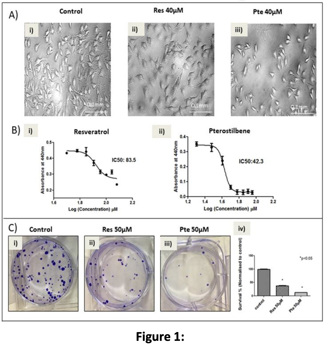
Figure 1: Pterostilbene is more potent in eliminating HeLa cervical cancer cells as compared to resveratrol: (A) Brightfield analysis of HeLa cells untreated (Ai) or treated for 24 h with 40 µM of resveratrol (Res; Aii) or 40 µM of pterostilbene (Pte; Aiii). Evidence of cell elimination was only seen robustly in cells treated with pterostilbene at 40 µM. (B) Analysis of IC50 values, generated by a
Pterostilbene Is More Potent in Eliminating HPV+ HeLa Cells Compared to Resveratrol
In order to study the comparative cytotoxicity of pterostilbene and resveratrol on HeLa tumor cells, brightfield images (Figure 1A) and WST-1 cell viability assays (Figure 1B) were performed 24 h post-treatment. The brightfield images taken after 24 h of treatment (Figure 1A) showed that pterostilbene (40 µM) eliminates significantly more cells than resveratrol at the same concentration. Live imaging of cells treated with 60 µM of the two compounds
Live
Of the two compounds, pterostilbene showed significantly more death and characteristic apoptotic blebbing with
.
Inhibition of Cell Migration of HeLa Cells Treated with Pterostilbene and Resveratrol
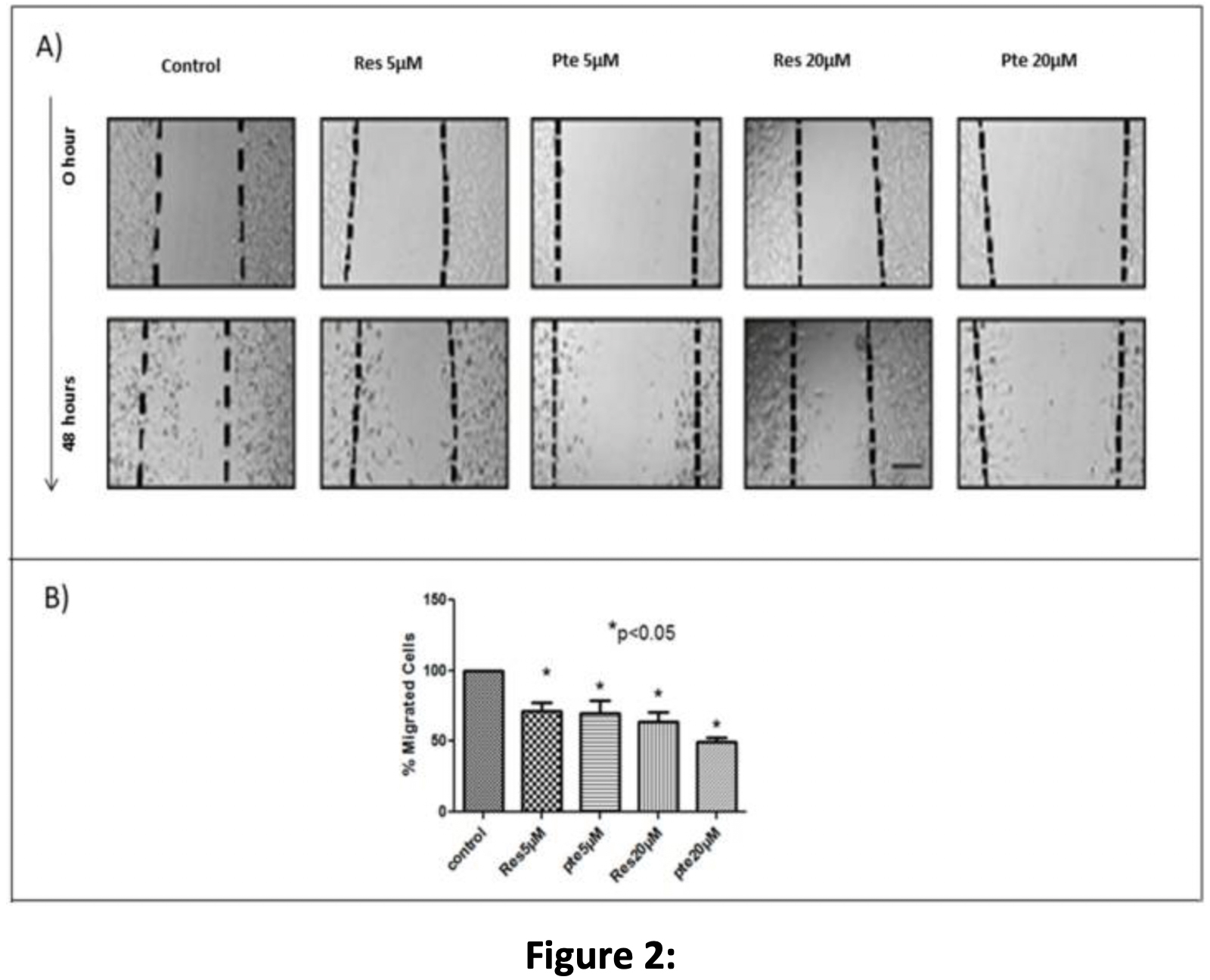
To determine the comparative efficacy of resveratrol and pterostilbene in inhibiting HeLa cell migration, two different sub-lethal concentrations of each compound were used in a 48-h scratch assay (Figure 2). Based on the WST-1 results and brightfield images (unpublished), we found that cells treated with a concentration below 25 µM showed no signs of cellular toxicity. To avoid any cytotoxicity, we used lower concentrations of 5 µM and 20 µM. At sub-lethal concentrations of 5 µM and 20 µM, both resveratrol and pterostilbene significantly inhibited HeLa cell migration relative to untreated cells (p < 0.05; Figure 2). Pterostilbene was more effective in inhibiting HeLa cell migration at 20 µM when compared to resveratrol; however, this result was
Figure 2: Resveratrol and pterostilbene inhibit cell migration: (A) HeLa cells were monitored for cell migration into a scratched “wound”. Cells were either untreated or treated with sub-lethal concentrations (5 µM and 20 µM) of resveratrol (Res) or pterostilbene (Pte). The extent of migration into the scratched area was calculated after 48 h and revealed that both resveratrol and pterostilbene significantly inhibit cell migration, although pterostilbene had
Cell Cycle Arrest at S-Phase in HeLa Cells Treated with Low Concentrations of Resveratrol and Pterostilbene
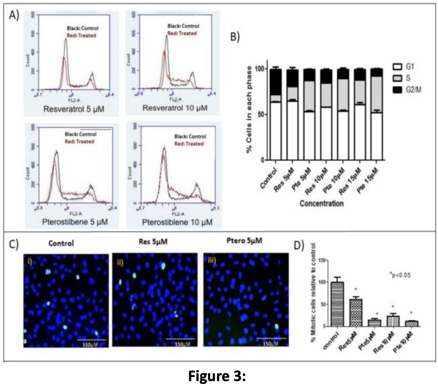
In order to compare the effect of sub-lethal doses of either resveratrol or pterostilbene on the cell cycle of HeLa cells, treatment was carried out with three different concentrations (5 µM, 10 µM, and 15 µM) of the two compounds for 18 h prior to flow cytometric analysis (Figure 3A). Flow cytometry analysis showed that the cells treated with either compound exhibited a significant decrease in the number of cells in the G2-M phase with respect to the control cells (p < 0.05) (Figure 3A
Figure 3: S-phase arrest in HeLa cells treated with low concentrations of resveratrol and pterostilbene: (A) Flow-cytometric evaluation of HeLa cells untreated or treated with sub-lethal doses of resveratrol (Res) and pterostilbene (Pte) for 18 h. Treated cells exhibited S-phase arrest and a subsequent decrease in the number of cells in G2/M. Pterostilbene was a more potent compound than resveratrol, showing a capacity to arrest cells at the S-phase at concentrations as low as 5 µM. (B) Graphical representation of the dose-dependent cell cycle effects induced by resveratrol and pterostilbene at three different concentrations (5 µM, 10 µM, and 15 µM). (B) The graph represents data from triplicate sample experiments normalized to the control (mean % cells in each phase ± S.E.M.) (C) Immunofluorescent images of HeLa cells probed for the M-phase marker
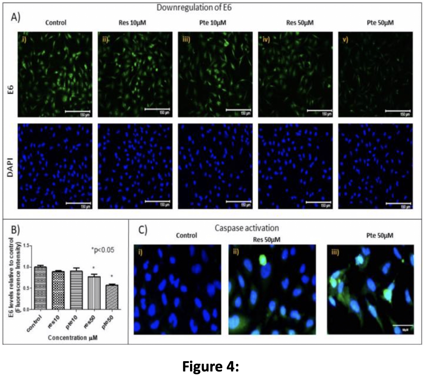
Downregulation of Viral Oncoprotein E6 and Upregulation of Active-Caspase-3 in HeLa Cells Treated with Pterostilbene and Resveratrol
In order to investigate how resveratrol and pterostilbene were affecting HeLa cell survival and cell cycle progression, we treated cells with either of the two compounds at sub-lethal (10 µM) and higher (50 µM) concentrations prior to analysis by immunostaining for E6, active caspase-3, and p53 (Figure 4A–C). At 10 µM, both resveratrol and pterostilbene failed to significantly affect levels of E6 and active caspase-3 levels relative to the control (Figure 4A
Figure 4: Downregulation of viral oncoprotein E6 and upregulation of active-caspase-3 in HeLa cells treated with resveratrol or pterostilbene: (A) HeLa cells immunostained for E6 levels (green) and counterstained with the nuclear dye 4’,6-diamidino-2-phenylindole (DAPI) (blue) after treatment with resveratrol (Res) and pterostilbene (Pte; 10 µM and 50 µM). Loss of E6 proteins
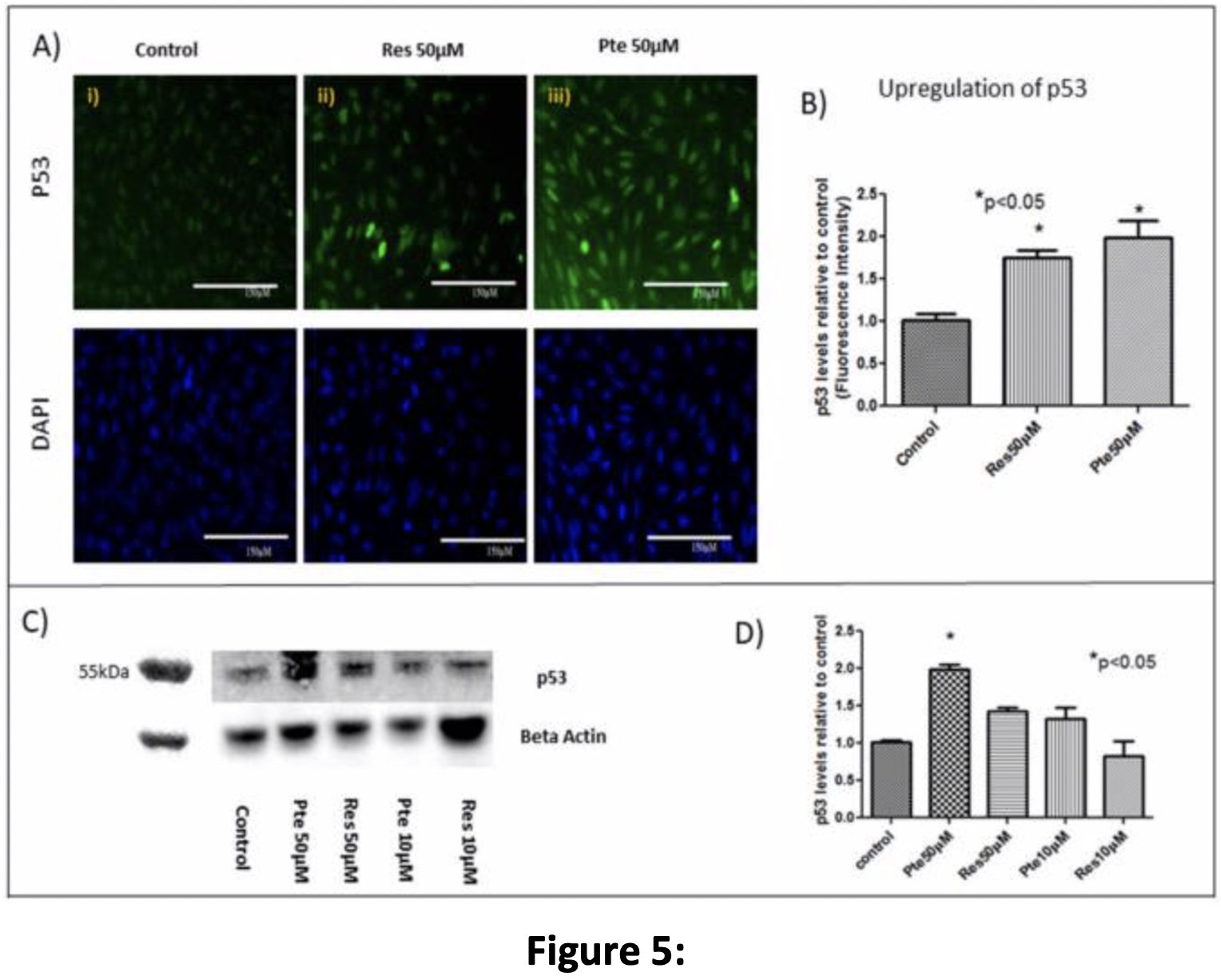
Upregulation of Tumor Suppressor Protein p53 in HeLa Cells Treated with Pterostilbene and Resveratrol
Concomitant with E6 suppression, 50 µM pterostilbene treatment for 22 h caused an upregulation of p53 in HeLa cells (Figure 5A
Conclusion
Cervical cancer is a major concern in developing countries due to
Here, we show that pterostilbene potently suppresses HPV E6 expression (Figure 4) and efficiently eliminates HPV+ cells in culture by p53-mediated apoptosis (Figure 1 and Figure 5) while suppressing cell proliferation (Figure 3) and migration (Figure 2). We find that pterostilbene is a more promising agent against cervical cancer when compared to resveratrol. Based on such properties, the use of pterostilbene presents a relatively economical but highly hopeful therapeutic approach to treat HPV infections and cervical cancers. Our future studies will include signaling studies using HPV+ murine tumor models to confirm these observations in vivo.